New approach to investigate the body’s binding between cells
The discovery of a new approach for the investigation of binding between cells allows for, in the longer-term, development of methods to identify and prevent the factors involved in the formation of cancer metastases.


The epithelium is a layer of cells that covers the outer surface of an organism, and as these play a major role in many aspects of medical science, many researchers study the epithelial cells. A research team from Aarhus University has decided to concentrate on examining the binding (adhesion) between epithelial cells and to determine the physical size of the connections that exist between epithelial cells. The binding between the cells is important in order to maintain the selective barrier that the epithelial tissue forms so that the body balance (homeostasis) is maintained.
Cancer cells from the epithelial tissue depend on being able to escape the bonds attaching them to the neighbouring cells in order to migrate and form tumors elsewhere in the body – a process called metastasis. It is therefore important to understand the bonds between cells, and what it takes for them to escape from one another. The researchers have now reached another important step on the way to understanding the bond between these cells. In the longer term, the researchers hope that their system can be used to study the factors involved in the formation of cancer metastases, so that it can be prevented.
The results have just been published in the internationally renowned journal Nano Letters by researchers from the Interdisciplinary Nanoscience Centre and the Department of Molecular Biology and Genetics at Aarhus University.
Description of the project
The epithelial tissue is located along all of the body’s surfaces and therefore constitutes the interface between the outside and inside. An important parameter for the maintenance of the epithelial tissue is that the epithelial cells form a selective barrier, which enables the cell to regulate which substances are allowed to pass the epithelial barrier and thus get in and out of the body.
The bonds between the cells consist of different cellular structures, including the "adherence junctions" consisting of a protein called E-cadherin, which can bind to other E-cadherin proteins in the neighbouring cells, whereby the cell-cell contact is established.
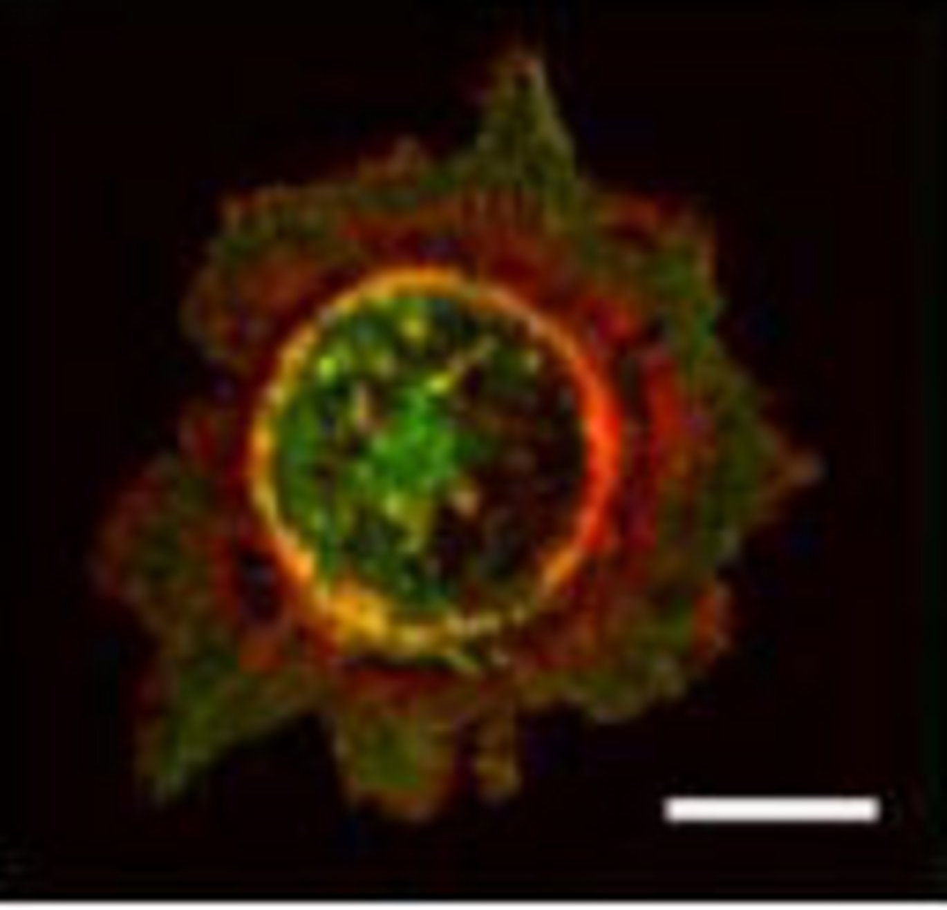
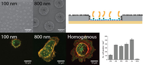
The researchers have studied the binding of epithelial cells to surfaces where the E-cadherin was coupled into circular nanostructures with a well-defined diameter. These surfaces were designed at the iNANO Centre using a technique known as "colloidal lithography".
Through coupling proteins to a modified, water-repellant gold surface, circular nanostructures of E-cadherin are formed and the surrounding regions are coated with a cell-repellent thin film. This ensures that the cells will only be able to establish contact with E-cadherin, which means that the researchers can study the binding after the formation of adherence junctions of varying sizes.
The nanostructures are thus used to simulate the epithelial membrane, and the cells growing on the surface will be presented with the E-cadherin in the same way and with the same density as if they met another epithelial cell.
By varying the size of the E-cadherin circles on the surfaces, the Aarhus researchers have been able to determine the minimum size of the adherence junctions required to maintain the bond between the cells. Their results showed that the minimum size of the adherence junctions was about 0.03 ?m2, which corresponded to an estimated number of about 17 E-cadherin proteins in each adherence junction. The system can be used to test what it takes to break this minimum bond, ie. when the cancer cells can escape from the neighbouring cells.
The project provides new basic knowledge for fundamental research on how a bond between cells is formed. The bond between epithelial cells is particularly relevant in cancer research in the metastasis of tumours, since a reduction of E-cadherin is known to be required for the cells to escape and start new cancer metastases.
Nano Letters ASAP (2012)
†Interdisciplinary Nanoscience Center (iNANO) and ‡Department of Molecular Biology and Genetics, Aarhus University, Denmark
More information
Lene Niemann Nejsum
Department of Molecular Biology and Genetics, Aarhus University, Denmark
nejsum@mb.au.dk, mobile: +45 21163121 (+45 87155492)
Duncan S. Sutherland
Interdisciplinary Nanoscience Center (iNANO, Aarhus University, Denmark
duncan@inano.au.dk, mobile: +45 23385789
http://inano.au.dk/nanobiointerfaces/
Text: Gitte A. Pedersen, Lene N. Nejsum and Lisbeth Heilesen
Translation: Lisbeth Heilesen
