
Zebrafish let you see the biological fate of nanoparticles in vivo
Ever wondered if you could see through the body of a living organism and observe the dynamic interplay between cells and nanoparticles injected into the bloodstream? This is now possible as the use of transgenic zebrafish embryos now offers a unique opportunity for intravital microscopy at imaging resolutions unrivalled by existing mammalian models.

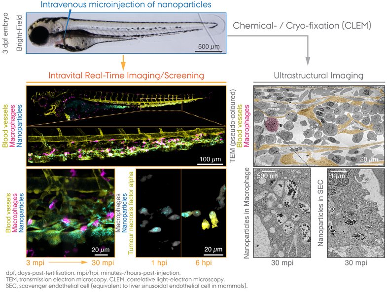
More than 99% of the total nanoparticle dose administered still fails to reach tumours – this shows a common challenge we face today for the development of targeted drug delivery. In particular, the liver is among the organs known to efficiently clear the blood of nanoparticles injected intravenously, a typical method of drug administration for systemic delivery. How this rapid blood clearance mechanism is achieved remains a "black box" as yet, in part due to the technical difficulties in bioimaging of nanoparticles at single-cell resolution. An international research team across Denmark, Germany and Italy has now established a "zebrafish way" of visualising the very processes in real-time and at ultrastructural resolution.
The research team focused on two cell types that are well-conserved across vertebrate animals: macrophages and scavenger endothelial cells. They are known as waste-removers in direct contact with the bloodstream, and the functional equivalent in our body are Kupffer cells and liver sinusoid endothelial cells, respectively, that reside at the blood-tissue interface of the liver. Transgenic zebrafish embryos can make them visible by means of genetically-encoded fluorescent proteins, and together with fluorescently-labelled nanoparticles the researchers revealed how circulating nanoparticles are almost exclusively captured by those cells within a time frame of several minutes to hours following injection into the blood. The real-time multicolour bioimaging strategy was further combined with electron microscopy approaches to enable (correlative) analysis of the tissue ultrastructures to unequivocally detect nanoparticles that are being taken up by endocytosis and subsequently localised in sub-cellular compartments.
These findings highlight the unique strength of a zebrafish model for genetic manipulation and intravital imaging that can deliver new exciting insights into the mechanisms of nanoparticle clearance once considered "invisible". Zebrafish thus now begin to emerge as an attractive tool to assist the development of bionanomaterials towards safer and more efficacious designs.
This study is a fruit of ongoing research led by Assistant Professor Yuya Hayashi at Aarhus University and his international collaborators Dr. Carsten Weiss and Prof. Uwe Strähle at Karlsruhe Institute of Technology (KIT), Germany. The collaboration was realised through two consecutive postdoctoral grants financially supported by Independent Research Fund Denmark | the Research Council for Technology and Production Sciences (DFF | FTP) and Lundbeck Foundation.
The Danish wing of the research was carried out with assistance of the zebrafish facility, headed by Dr. Kasper Kjær-Sørensen and Prof. Claus Oxvig at Department of Molecular Biology and Genetics, Aarhus University.
The article was published in ACS Nano, a highly renowned journal in Nanoscience.
"Differential Nanoparticle Sequestration by Macrophages and Scavenger Endothelial Cells Visualized in Vivo in Real-Time and at Ultrastructural Resolution" by Yuya Hayashi*, Masanari Takamiya, Pia Bomholt Jensen, Isaac Ojea-Jiménez, Hélicia Claude, Claude Antony, Kasper Kjær-Sørensen, Clemens Grabher, Thomas Boesen, Douglas Gilliland, Claus Oxvig, Uwe Strähle, and Carsten Weiss.
ACS Nano 14 (2020) pp. 1665-1681. https://doi.org/10.1021/acsnano.9b07233.
Movie (see the movie)
Macrophages (magenta) with internalised nanoparticles (cyan) crawling along the inner side of blood vessels (yellow). Tg(fli1a:eGFP); Tg(mpeg1:mCherry) embryos at 3 dpf were injected with Pacific Blue-labelled 70 nm SiO2 nanoparticles (2 ng). Time-lapse imaging was performed at the intervals of every 16 s for 15 min at 1-4 hpi. (Reprinted from Hayashi et al. (2020) ACS Nano. Copyright 2020 American Chemical Society)
For further information, please contact
Assistant Professor Yuya Hayashi
Department of Molecular Biology and Genetics
Aarhus University, Denmark
yuya.hayashi@mbg.au.dk