
How does a lizard lose its tail?
Researchers at Aarhus University have now found the answer to this question, attracting enormous international attention.
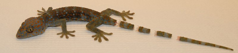
When a lizard feels threatened by a predator, it can shed its tail at will. Because the tail maintains the ability to move up to half an hour after separation, the lizard hopes that the predator will attack the wriggling tail, enabling it to make a getaway in the meantime.
However, there are certain disadvantages associated with this ability, as the tail is used for purposes such as storing fat. It is therefore an advantage for the lizard that it only sheds a minor portion of its tail. This self-amputation (autotomy) of the tail follows a determined pattern, as the tail has pre-formed ‘dotted lines’. The researchers have now described these perforations in such detail that it is possible to explain the autotomy mechanism.
They hope that this discovery can inspire material scientists and engineers in their design of new materials, where quickly detachable structures are important.
Studies in autotomy
The project involved studies of what makes a lizard capable of shedding its tail. The researchers showed that there are pre-severed ‘score lines’, and they used advanced bio-imaging techniques to characterise the structure of these lines.
Making the discovery actually happened quite by chance. The research team is headed by Professor Jan J. Enghild, Aarhus University, and the group is interested in areas such as identifying new proteolytic enzymes (enzymes that break down other proteins). In addition to their basic fascination with autotomy, the researchers are interested in identifying new enzymes, and this was the starting-point for the project that has now been published.
“We imagined that proteolytic enzymes might be involved in the process, partly because we considered there would be a breakdown of tissue, and partly because such a breakdown is normally carried out by proteolytic enzymes. If this was the case, there had to be incredibly efficient and aggressive enzymes that could be worth taking a closer look at, as they could potentially be used in industry,” says Professor Enghild.
In connection with the study, the team used a modern method (proteomics) to identify proteins – and thereby enzymes as well – in complex combinations. This study showed that proteolytic enzymes are not involved in autotomy.
Pre-formed fracture surfaces identified
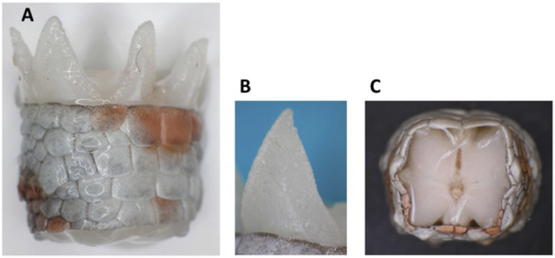
The new discovery motivated the researchers to take a closer look at the lizard’s fracture surfaces. They therefore teamed up with Aarhus University colleagues in biology, medicine and chemistry, who analysed the surfaces using conventional cross-section tissue samples, as well as modern methods such as scanning electron microscopy (SEM) and magnetic resonance imaging (MRI), all of which were at the disposal of the very cross-disciplinary project group.
After the team had collected and compared the results from the different methods, it became more and more clear for them that autotomy is a mechanical process rather than a biochemical one. There are pre-formed fracture surfaces at determined intervals right along the lizard’s tail, and the new data indicate that the structure of the muscle fibres in the surface changes in connection with autotomy, whereby the interaction between the fracture surfaces is weakened and autotomy can take place.
It is many years since a thorough study of autotomy was made, and the modern equipment used in this study was not available at that time.
“It’s the new methods and cross-disciplinary approach to the issue that’s made it possible for us to dig deeper into an understanding of autotomy,” says Kristian W. Sanggaard, a member of the project team.
The article has been published in PLOS ONE, and includes an illustrative 3D film of the tail based on the MRI results.
For more information, please contact
Kristian Wejse Sanggaard
Department of Molecular Biology and Genetics and
Interdisciplinary Nanoscience Centre (iNANO)
Aarhus University
+45 2374 1497
krs@mb.au.dk