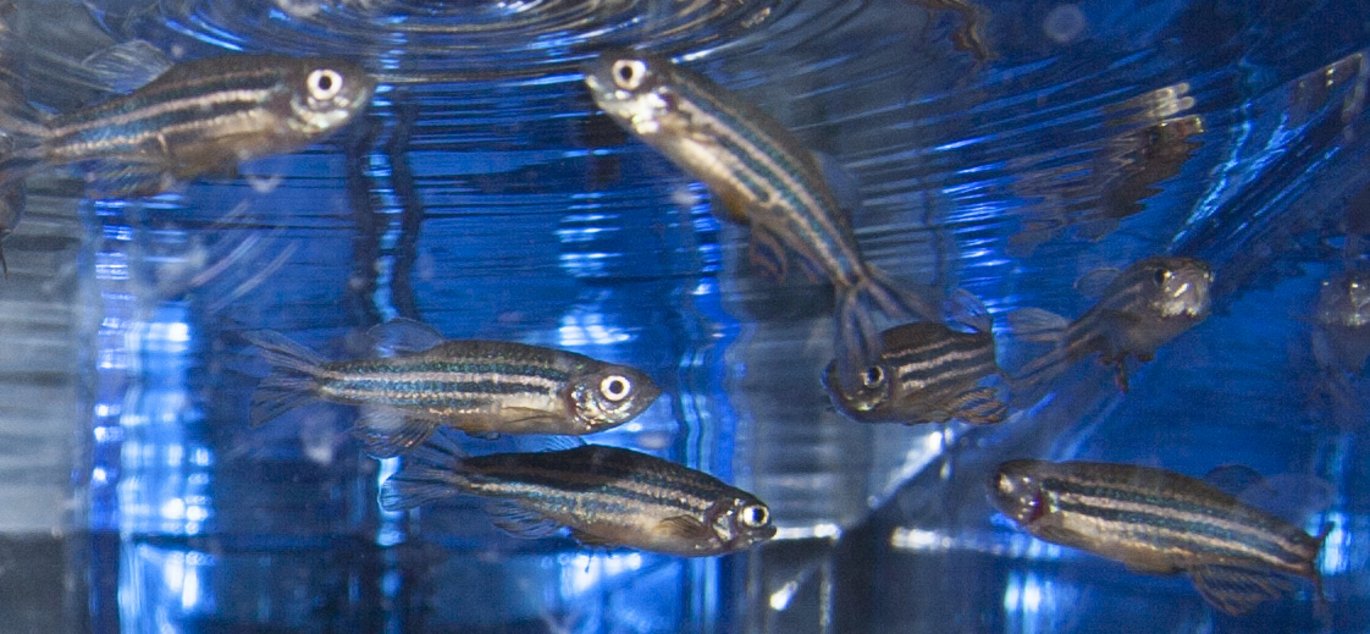
The zebrafish is an important animal model
Humans and zebrafish have more in common than you would think. Therefore, zebrafish are used more and more, for example, to study the function of genes, to create animal models for human diseases and to develop new human drugs.


At first, it may seem strange that an ordinary Danish aquarium fish has become the researchers' new favorite. Nevertheless, this is the case for the small zebrafish (Danio rerio), which is currently used for studying the function of genes to create animal models for human diseases and to develop new human drugs. We have much more in common with the zebrafish than you might think, and zebrafish are naturally equipped with a number of properties that make them very suitable in the research laboratory.
The zebrafish is similar to humans anatomically and genetically
From the outside, the similarities between humans and zebrafish are limited, but with a few exceptions – such as mammary glands, placenta and lungs – virtually all human organs are also found in zebrafish. Generally, the organs consist of the same cell types as in humans, and in the cells, largely the same genes dictate the identity and function. For 71 percent of all human genes, a corresponding zebrafish gene has been identified, and when looking at genes associated with human disease, the number is as high as 80 percent. Not surprisingly, the mouse is more similar to us, as there are mouse versions of 83 percent of the human genes, whereas in chicken is it 64 percent.
Useful characteristics of the zebrafish
Some of the characteristics that make the zebrafish well suited as an animal model are its high fertility, fast embryonal development outside the parent fish and a transparent embryo that allows for cell development to be examined from the outside. Assuming optimal environmental management, water quality and health, a single pair of zebrafish can produce 200 fertilized eggs by just a single weekly crossing, which can take place every week all year round.
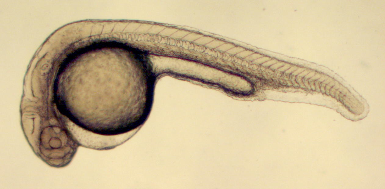
In the days leading up to the crossing, males and females are kept separated. When the females and males are together again, the females spawn within ten minutes, and the males fertilize the eggs immediately. The fact that the zebrafish eggs are fertilized outside the mother, and that the embryo remains transparent for several days, means that each cell can be monitored continuously in real time by ordinary light microscopy within the living embryo.In addition to the embryonic development, the transparency also enables detailed studies of the early larval stages.This is a huge advantage as this means that the phenotypes – which might not be readily visible in, for example, a mouse – can be detected. There are also pigment-free zebrafish lines which allow light microscopic studies of the inside of even the adult fish, although the size, of course, limits the light penetration into the underlying tissue.
Manipulation of gene function in zebrafish
In order to study the function of a gene, the activity of the gene or its products (RNA, proteins) is manipulated. With the technology available in zebrafish, it is possible to turn off or turn down a specific gene, to increase the gene or even introduce foreign genes.
It is also possible to create specific mutations and other changes in the genome exactly where you want.
Zebrafish in clinical research
In addition to basic research into the function of genes and developmental biology, zebrafish are used to study the genetic basis of a wide range of human diseases - such as cardiac arrhythmias, neurodegenerative diseases, endocrinological disorders and hematological disease.
In addition, zebrafish are also used in studies of the genetic and neurological basis of depression, drug addiction and learning.
Also, the zebrafish is becoming a recurring model animal in studies where large libraries of low molecular weight substances are screened for medicinal properties against specific diseases or disease processes.
Screening of new drugs
A major reason for the use of zebrafish in drug screening is the availability and size of the embryos, and the fact that the embryos can absorb the test substances directly from the water. The small size means that embryos can be kept in very small volumes, even in 96-or 384-well ELISA trays.
Therefore, large libraries of test compounds can be screened in a living organism in a very manageable format, and the consumption of expensive test substances is minimal.
Toxicological studies
By screening new drugs in a living organism, you get information about the pharmacological effects as well as important knowledge about possible toxic effects: Do the embryos survive, is the heart function affected, is hepatic necrosis induced or is the respiration affected? Such information is not obtained in experiments with cultured cells, and the information is considerably more expensive and often more complex to achieve in mammals. Therefore, the zebrafish is an ideal supplement, where toxic effects are examined in zebrafish embryos before they are tested on rodents and larger mammals.
The zebrafish as a supplement to other model organisms
The zebrafish also has great research value as a supplement to other model organisms. An example of a beneficial synergy is that zebrafish can be used to screen for phenotypic changes resulting from specific genetic manipulation relatively quickly and inexpensively at high resolution. This data can then be used to guide the phenotyping of mice with the same gene alteration, and thus zebrafish can be used to guide future experiments on mammals. It has been possible to identify phenotypes in zebrafish, which otherwise had been overlooked in mice. Also, data from mice can also be followed up by high-resolution real-time analysis of dynamic processes in zebrafish, such as cell migration, angiogenesis or axonal outgrowth.
The zebrafish as a supplement to other model organisms
The zebrafish also has great research value as a supplement to other model organisms. An example of a beneficial synergistic is that zebrafish can be used to screen relatively quickly, cheaply and with a high resolution for phenotypic changes resulting from specific genetic manipulation. This data can then be used to guide the phenotyping of mice with the same gene alteration, and thus zebrafish can be a springboard for future experiments on mammals.
Thus, it has been possible to identify phenotypes in zebrafish, which otherwise had been overlooked in mice. Also, data from mice can also be followed by high-resolution real-time analysis of dynamic processes in zebrafish, such as cell migration, angiogenesis or axonal outgrowth.
Housing of zebrafish
As the zebrafish is a schooling fish, they are kept in groups with a recommended density of up to five adult fish per liter. This allows them to live out their natural schooling behavior. In the standard configuration of a standard commercial aquarium system for zebrafish, there is room for more than 900 adult fish in an area corresponding to a standard mouse shelf for 75-150 mice.
This helps to make zebrafish relatively inexpensive to work with, and you therefore get more research for less money.
The embryonic development of the zebrafish
The embryonic development of the zebrafish is extremely fast: approx. 3 hours after fertilization the embryo has 1,000 cells, after about 18 hours after fertilization, the embryo begins to move, and about 22 hours after fertilization, the heart beats.
For further information, please contact
Kasper Kjær-Sørensen, PhD
Department of Molecular Biology and Genetics
Aarhus University
kks@mbg.au.dk - +45 5144 6497
Claus Oxvig, Professor
Department of Molecular Biology and Genetics
Aarhus University
co@mbg.au.dk - +45 3036 2460
Aage Kristian Olsen Alstrup, PhD, Veterinarian
Department of Clinical Medicine, the PET Centre
Aarhus University
aage.olsen@clin.au.dk - +45 78464396
This article was published in Dyrlægen (The Veterinarian) on 27 February 2017, pp. 26-30, and is reproduced here by permission of the editor of Dyrlægen Dyrlægen.
