The Interferon group
The research projects in the interferon group are centered around the cellular function of two families of interferon induced genes, ISG12 and the 2-5A system. In addition, we are engaged in uncovering a signal transduction pathway initiated by the vitamin folic acid leading to activation of the oncogene STAT3. And finally, we are studying the disease endometriosis with focus on the infertility accompanying this disorder.
The tecniques employed in the research projects cover mammalian cell culture and baculovirus expression combined with molecular biological analyses of proteins, RNA and DNA as well as analyses of tissue samples.
This project focuses on how the type 1 interferon controlled family of Interferon Stimulated Gene 12 (ISG12) proteins are involved in regulation of programmed cell death (apoptosis). Apoptosis is a morphological distinct form of programmed cell death essential for normal development and tissue homeostasis. Overexpression of individual members of the ISG12 proteins lead to apoptosis in different cell types in culture.
In order to establish the importance of ISG12 for apoptosis during embryonic development, we want to determine, which cells express ISG12 during early development. As model systems, we have chosen zebrafish, mice and nematodes. Since ISG12 encoding genes have been found in zebrafish D. renio and mice, it is possible to knock down ISG12 expression and in this way uncover the importance of ISG12 during early development. These studies will help eludidating if ISG12 plays a fundamental role in apoptosis during early development as well as in the cellular apoptotic network.
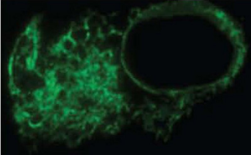
Mitochondria produces cell energy, however, mitohondria also plays a very important role in apoptosis, as the outer mitochondrial membrane is permeabilized during most apoptotic pathways. We have found that ISG12 is localized to the inner mitochondrial membrane, but unknown localization signals locate ISG12 here. We want to identify the peptide sequences in ISG12 responsible for this cellular localization. The identified localization signals can putatively be used for targeting specific proteins and drugs to the inner mitochondrial membrane. This can ultimatively be explored for drug development targeting the mitochondria.
The expression of 2’-5’-Oligoadenylate synthetase (OAS) is induced by Type 1 interferons in response to viral infection. The OAS proteins have a unique ability to produce 2’-5’ Oligoadenylates (2-5A), which bind and activate the ribonuclease RNase L. The RNase L degrades cellular RNAs which in turn inhibits protein translation and induces apoptosis. The level of 2-5A is regulated by synthesis by the 2’-5’Oligoadenylate Synthetases (OAS) and by degradation by 2’ phosphodiesterases (2’PDE). This project is centered around the characterization of the enzymes OAS and PDE with focus on OAS in primitive organisms and PDE in human cells.
We have found that many primitive organisms have an OAS gene wih a function different from the function in the 2-5A system. Furthermore, we found that the specific 2’PDE named PDE12 and one of the OAS1 isoforms are localized in the mitochondria, and as a consequence a full 2-5A system can now be attributed to the mitochondria. Specifically, we have shown that PDE12 is involved in the degradation of mitochondrial mRNA.
It has previously been shown that polymorphisms in the human OAS1 gene are correlated with diseases like diabetes. The specific localisation in the mitochondria that we found of the OAS1 isoform is linked to these polymorphisms. We are investigating how expression of specific OAS isoforms are involved in the development of diabetes.
The tumour promoting properties of high folate diet has up till now been overlooked. More focus is presently being put on the consequences of the folic acid fortification throughout the world and in particularly in USA. Ingestion of high doses of folic acid through fortified food and vitamin supplements might lead to increased growth of cancer cells with Folate Receptors on the cell surface. We have identified a novel signal transduction pathway induced by folic acid in cells in culture. Our current research focuses on how to inhibit cancer cell proliferation and the activation of the oncogene Signal Transducer and Activator of Transcription 3 (STAT3) via the cancer-specific Folate Receptor. Further research aiming at uncovering the molecular mechanisms involved in this signalling pathway, as well as solving the crystal structure of the Folate Receptor might pave the way for an intelligent drug design of small molecules acting as Folate Receptor antagonists
Endometriosis is a chronic disease in which endometrium-like tissue is located outside the uterus, frequently in the pelvic cavity but also in more distant regions. The symptoms include subfertility, chronic pelvic pain, dysmenorrhoea (painful periods), and dyspareunia (pain during intercourse). Today endometriosis can only be diagnosed either by ultrasound if the ovaries are involved or by operation. It has been estimated that 5 to 10% of fertile women are suffering from the disease and in a population of women with dysmenorrhoea, around 50% have endometriosis. Around 40% of women attending a fertility clinic have endometriosis. The pathogenesis of endometriosis is unclear. The risk is increased 7-9-fold for women who have a close relative (mother and/or sister) with endometriosis, indicating some genetic involvement. This project focuses on the decreased fertility in women with endometriosis. We study the molecular characteristics of implantation, in particular if either interferon, folic acid, or expression of specific homeobox proteins are involved in the decidualization process, which is the preparation of the endometrium for implantation. This knowledge can be used for improving the success of implantation after in vitro fertilization (IVF) both in women with endometriosis as well as in infertile women without endometriosis.